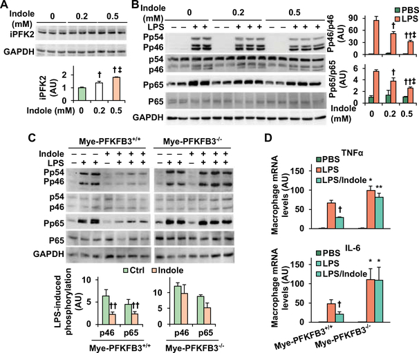
Figure 5. Indole stimatules PFKFB3 expression and suppresses macropahge proinflammatory actiaiton in a PFKFB3-dependent manner.
(A) Indole increases iPFK2 amount. (B) Indole suppresses macrophage proinflammatory signaling. For A and B, RAW264.7 cells were treated with or without indole (0.2 or 0.5 mM) for 24 hr in the absence or presence of LPS (100 ng/mL) for the last 30 min. (C,D) PFKFB3 disruption blunts the effect of indole on suppressing macrophag proinflammatory activation. Bone marrow cells were isolated from Mye-PFKFB3–/– and Mye-PFKFB3+/+ mice and differentiated into macrophages (BMDM). After differentiation, BMDM were treated with indole (0.5 mM) for 24 hr in the absence or presence of LPS (100 ng/mL) for the last 30 min (C) or LPS (20 ng/mL) for the last 6 hr (D). For A - C, cell lysates were subjected to Western blot analysis. Bar graphs, quantification of blots. For A - D, numeric data are means ± SEM. n = 4 – 6. *, P < 0.05 and **, P < 0.01 Mye-PFKFB3–/– vs. Mye-PFKFB3+/+ with the same treatment (LPS or LPS/Indole) (in D); †, P < 0.05 and ††, P < 0.01 Indole vs. Ctrl (in the absence of indole) (in A) under LPS-treated condition (in B) for the same protein (in C) or LPS/Indole vs. LPS within the same genotype (in D); ‡, P < 0.05 Indole (0.5 mM) vs. Indole (0.2 mM) (in A) under LPS-treated condition (in B).

