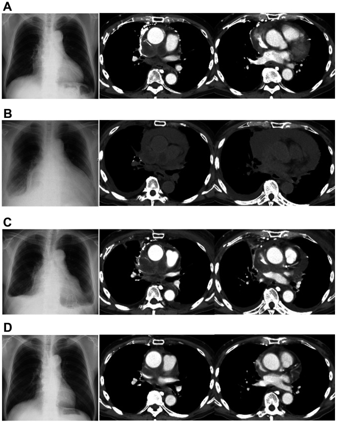
Figure 2.
Changes in imaging of the patient over time. (A) Before treatment with S-1, a chest radiograph revealed mild enlargement of the cardiac silhouette. Chest computed tomography demonstrated mediastinal tumor and minor amount of pericardial effusion. (B) Chest radiograph showing apparent enlargement of the cardiac silhouette and pleural effusion. Chest computed tomography revealed apparent enlargement of mediastinal tumors and an increase in pericardial effusion. (C) After drainage, pericardial effusion decreased but was still present. (D) Mediastinal tumor and pericardial effusion markedly decreased after combination therapy of carboplatin and etoposide was initiated.

