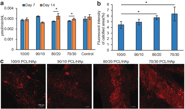
FIG. 7.
Assessment of mineralization and calcium deposition of osteodifferentiated cultured cells on the PCL/HAp samples. (a) Alkaline phosphatase activity of osteoblasts on days 7 and 14 postdifferentiation, which shows that cells are differentiating; also, increasing HAp in groups cells are more directing to differentiation (n = 3 per group; α = 5%). (b) Quantified fluorescence intensity (n = 3 per group; α = 5%) and (c) confocal images of xylenol orange staining of calcium deposition of differentiated cells at day 35 postdifferentiation, which shows significant increase in groups with higher concentration of HAp.

