
Fig. 1.
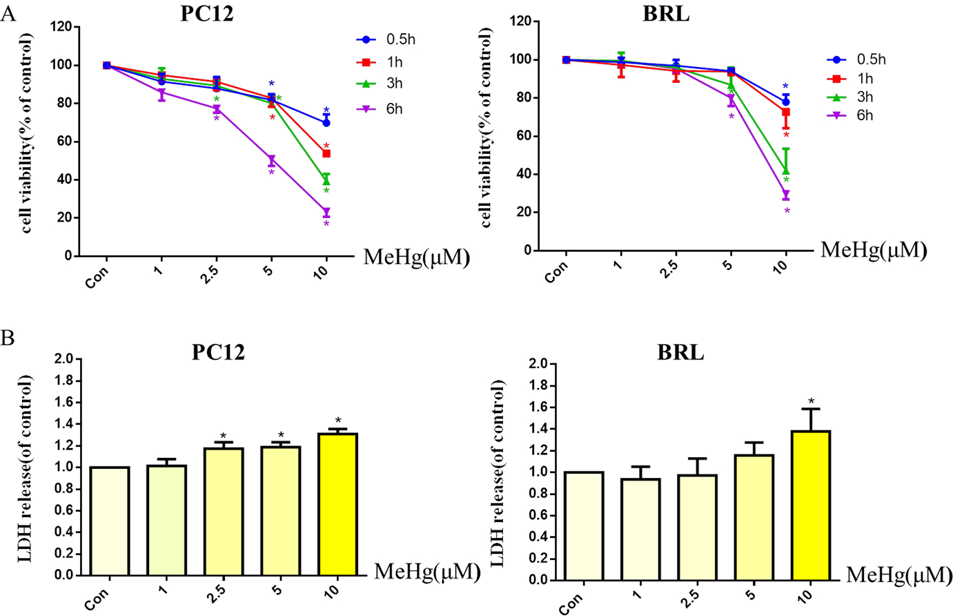
Effects of methylmercury (MeHg) on cell viability and cytotoxicity in PC12 and BRL cells. PC12 and BRL cells were treated with various concentrations (1–10 μM) of MeHg for 0.5 to 6 h. Cell viability (A) was measured by the MTT assay and cytotoxicity (B) was detected via lactate dehydrogenase (LDH) release. Data show mean ± standard deviation (n = 3).
*p < 0.05, compared with the control group. Statistical analysis was performed by one-way analysis of variance followed by a Dunnett test, a multiple comparison procedure.