Fig. 2.


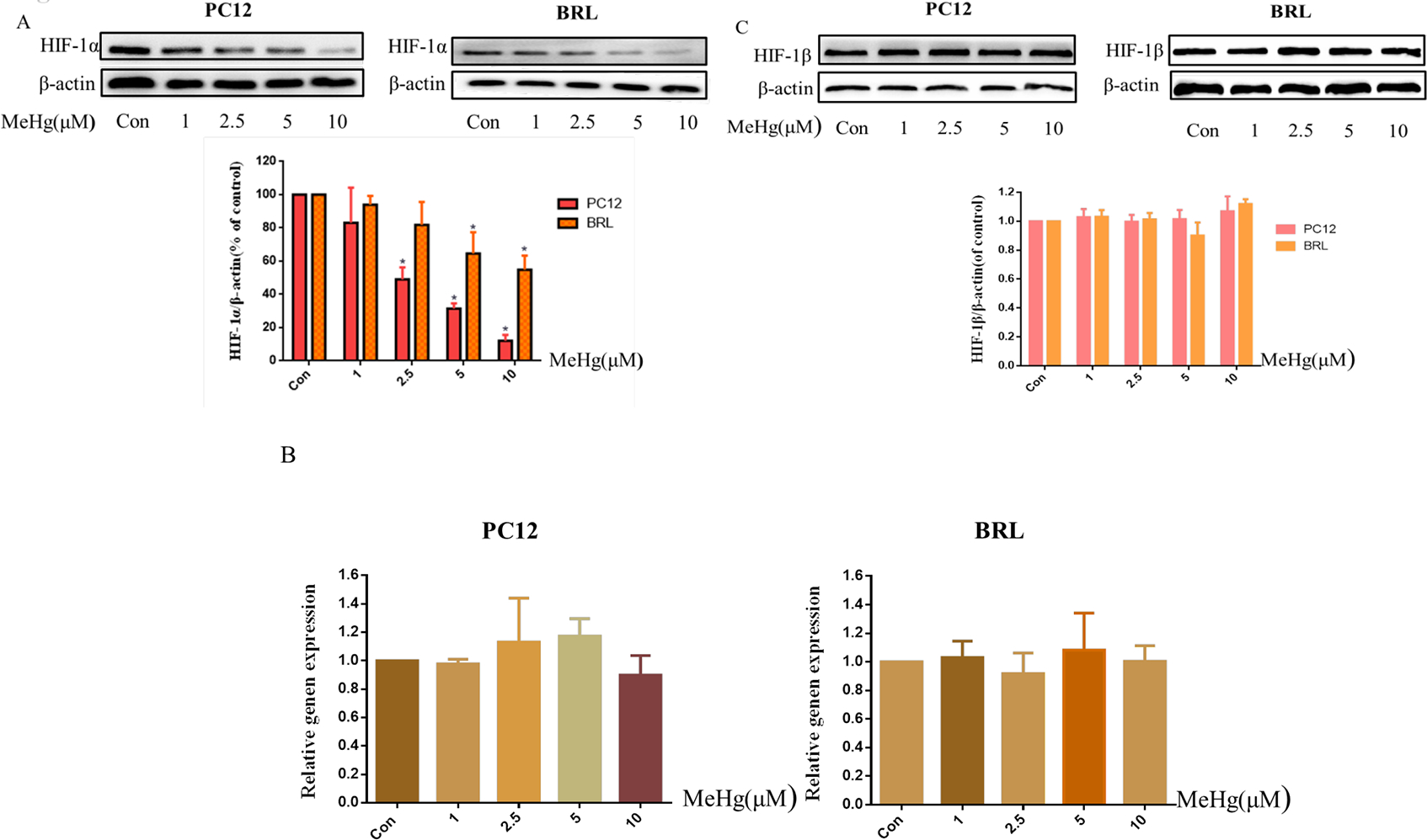
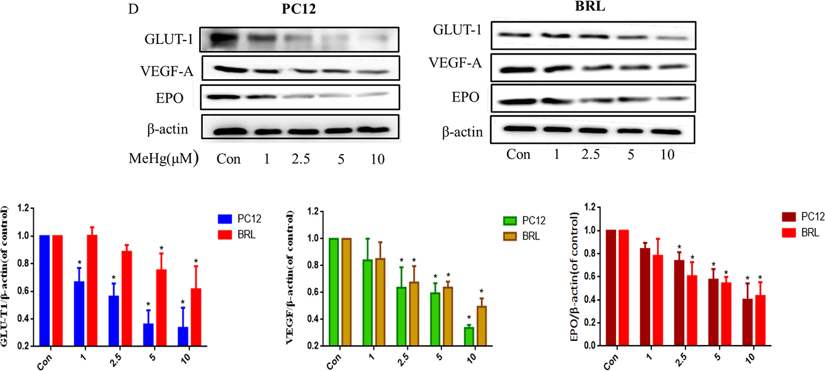
Protein levels of HIF-1α and its downstream targets in PC12 and BRL cells after treatment with methylmercury (MeHg). A) Western blotting of HIF-1α in cells treated with different concentrations (1–10 μM) of MeHg for 0.5 h. B) Quantitative RT-PCR was used to measure HIF-1α mRNA expression. C) Western blotting was used to detect the protein level of HIF-1β. D) The downstream proteins of HIF-1α glucose transporter-1 (GLUT-1), vascular endothelial growth factor-A (VEGF-A), and erythropoietin (EPO) after exposure to various concentrations (1–10 μM) of MeHg. β-Actin served as a loading control. Data show mean ± standard deviation (n = 3).
*p < 0.05, compared with the control group. Control groups were treated with free media without any agents. Statistical analysis was performed by one-way analysis of variance followed by a Dunnett test, a multiple comparison procedure.
