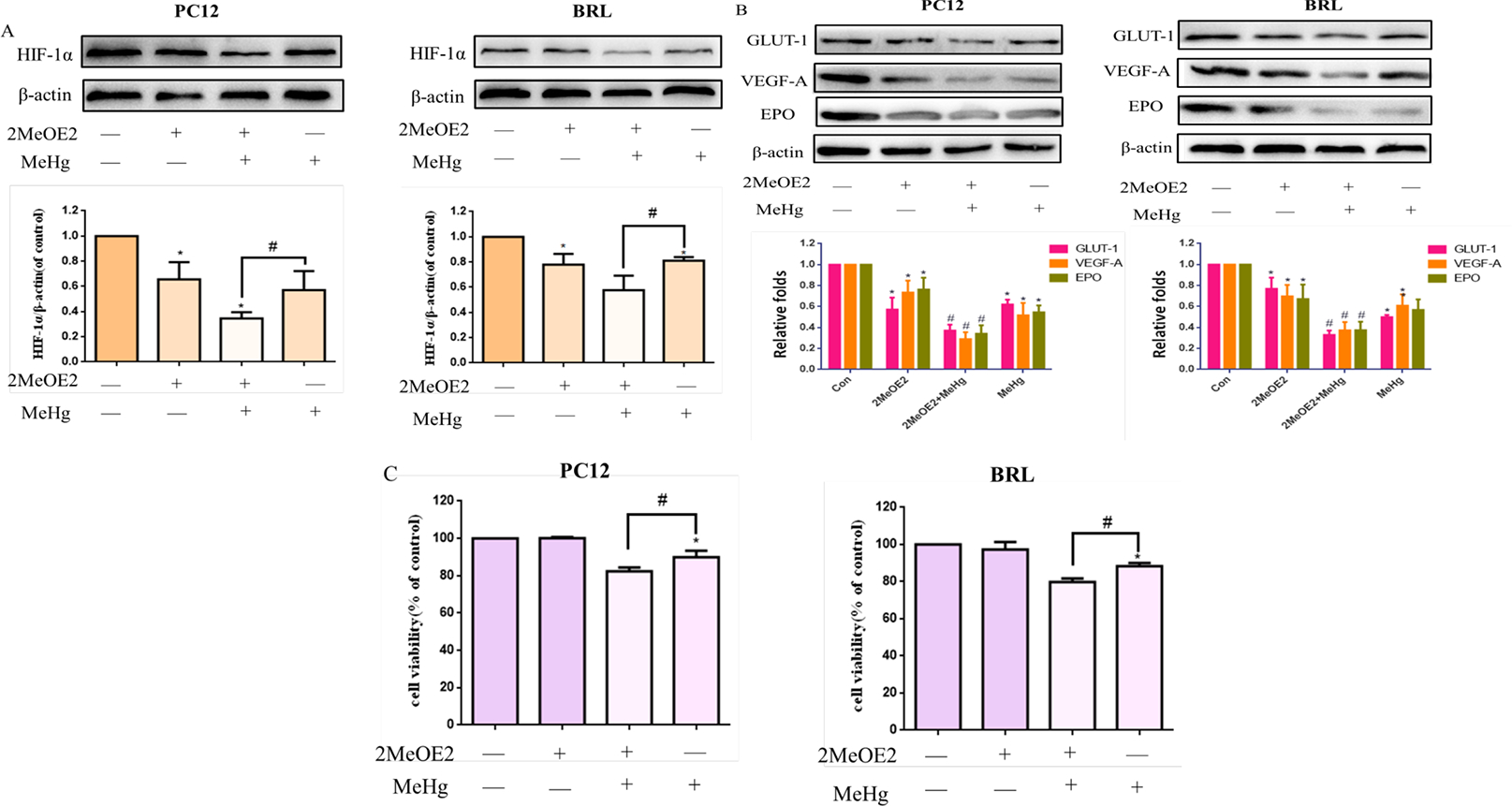
Fig. 7.

Effects of 2-methoxyestradiol (2-MeOE2) on methylmercury (MeHg)-induced acute cell injury. PC12 and BRL cells were pretreated with 2-MeOE2 (10 μM for 0.5 hours), followed by treatment with MeHg. A, B) Protein expression of HIF-1α and downstream targets glucose transporter-1 (GLUT-1), vascular endothelial growth factor-A (VEGF-A), and erythropoietin (EPO) was analyzed by western blotting. β-Actin served as the loading control. C) Cell viability was measured by MTT assay. *p < 0.05, compared with the control group; #p < 0.05, compared with the MeHg group. Control groups were treated with free media without any agents. Statistical analysis was performed by one-way analysis of variance followed by a Dunnett test, a multiple comparison procedure.
