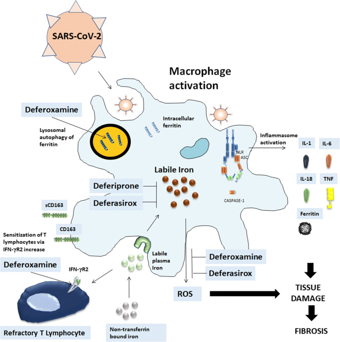
Fig. 1.
Iron chelation therapy in SARS-CoV-2 infection. SARS-CoV-2, likely through inflammasome activation, leads to stimulation of infiltrating macrophages that can promote hyperinflammation, characterized by increased levels of IL-6, TNF-α, IL-1β, ferritin and subsequent possible lung fibrotic complications. The increased ferritin production allows adequate storage of iron and deprives the pathogen of iron. Labile iron in the cell contributes to the formation of reactive oxygen species that further promote tissue damage and fibrosis. Iron accumulates in the reticuloendothelial macrophages and the shedding of CD163 is the marker of macrophage activation. Iron chelation therapy can interrupt these steps. (a) Deferoxamine (DFO) has a direct effect on ferritin since promotes ferritin degradation in lysosomes by inducing autophagy. Both deferiprone and deferasirox are likely to chelate cytosolic iron and iron which is extracted from ferritin prior to ferritin degradation by proteasomes. (b) DFO can induce an up-regulation of IFN-γR2 expression on the cell surface on activated T cells thus restoring T cell response to SARS-CoV-2 infection. (c) Deferasirox and DFO reduce fibrosis-inhibiting the production of free radicals, macrophage tissue infiltration and cause a remarkable decrease of IL-6 levels

