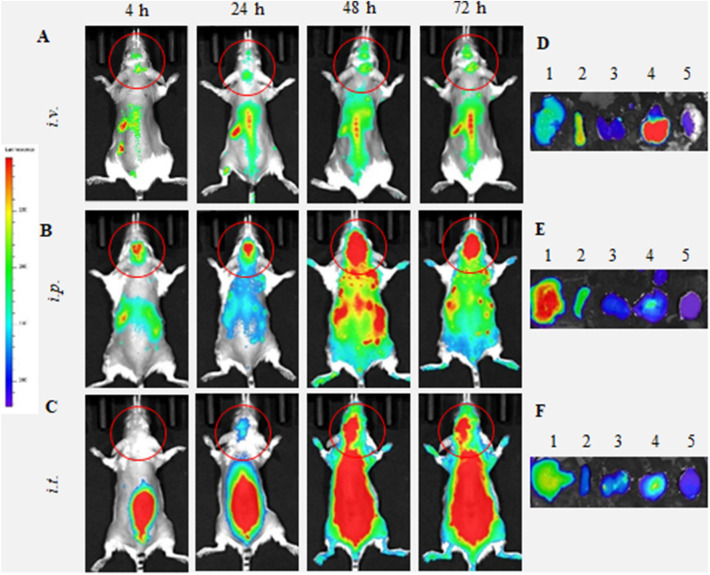
Figure 1.
Biodistribution of DIR-labeled BMM in Parkin Q311(X)A mice by IVIS. DIR-labeled BMM were administered in PD mice (12 mo. of age) (A, D) i.v. (4 × 10⁶ cells/200 µL), (B, E) i.p. (4 × 10⁶ cells/200 µL), or (C, F) i.t. (1 × 10⁶ cells/50 µL). Images were recorded (A–C) in live animals at different time points, and (D–F) in main organs, liver (1), spleen (2), kidney (3), lungs (4), and brain (5), collected at the endpoint (72 h). Prone representative images show DIR signal accumulation in the brain for all administration routes examined, especially at 24–72 h. Accumulation of labeled macrophages was also observed in the main peripheral organs.

