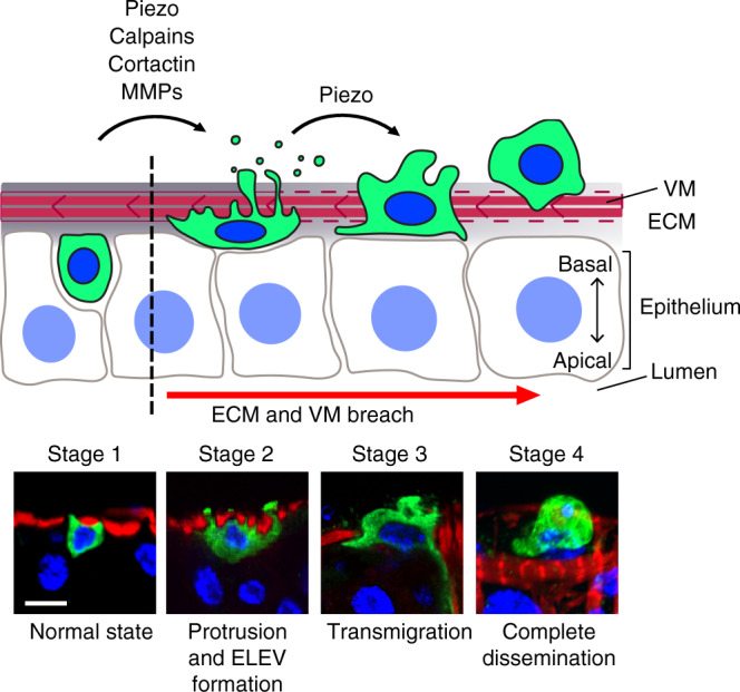
Fig. 7. Schematic stages of cell dissemination.

Stages of cell dissemination were reconstituted based on confocal images of fixed RasV12 cells and ex vivo live imaging of esgts > RasV12 posterior midguts. Lower panels show representative confocal images of control (esgts; stage 1) and RasV12 cells (stages 2–4). An esgts cell is illustrated in stage 1. In the absence of RasV12 expression, ISCs and EBs reside within the midgut epithelia. Upon RasV12 expression, invasive protrusions are formed at the basal side of the cells, and Mmp1 levels increase. Stage 2 illustrates a RasV12 cell producing large protrusions/blebs and ELEVs across the VM. These cells could be observed at days 2 and 3 of RasV12 expression. Stages 3 and 4 illustrate RasV12 cells under and after transmigration, respectively. Disseminated cells were frequently detected at days 2 and 3 of RasV12 expression. Scale bar, 10 µm.
