Figure 3.
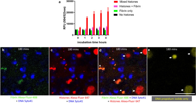
Protection of neutrophils from histone cytotoxicity by fibrin. (a) Sytox Green neutrophil viability assays were performed as in Fig. 2, with fibrin (as a suspension of FDP) in place of fibrinogen. Fibrin protected cells from the cytotoxic effects of histones. Error bars show 95% CI for the mean, n = 3. (b-e) Live cell microscopy studies were performed by adding fibrin (derived from Alexa-Fluor 488-labelled fibrinogen in green) and Alexa-Fluor 647 labelled histones (red) to neutrophils in a glass-bottomed dish. Images were taken after 180 min of incubation. (b) fibrin and (c) histone fluorescence with (d) as the merged image to highlight complexes between fibrin and histones (arrow heads). (e) Is the propidium iodide signal only. White arrows in (d) and (e) highlight some binding of extracellular histones to DNA released from damaged cells. The scale bar is 25 µm. Representative images are shown from 1 of n = 3 independent experiments.

