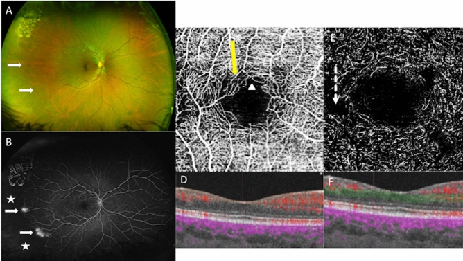
Figure 3.
Multimodal imaging of the right eye of a 30-year old woman with Sickle cell retinopathy (SS), stage 3. (A) Red/green ultra-wide field retinography with neovascular lesions (white arrows). (B) The ultra-wide field fluorescein angiography shows temporal peripheral non-perfusion area (white stars), and preretinal neovascular lesions (white arrows). (C) OCT angiography shows microvascular abnormalities in the perifoveal area in the superficial capillary plexus: areas of rarefied capillary (yellow arrow), and disruption of the perifoveal anastomotic capillary arcade (white arrow-head). (D) B-scan with the limits in orange of the SCP segmentation. Blood flow appears in red. (E) OCT angiography shows microvascular abnormalities in the perifoveal area in the deep capillary plexus: enlargement non-perfusion area (dotted arrow). (F) B-scan with the limits in green of the DCP segmentation. Blood flow appears in red.

