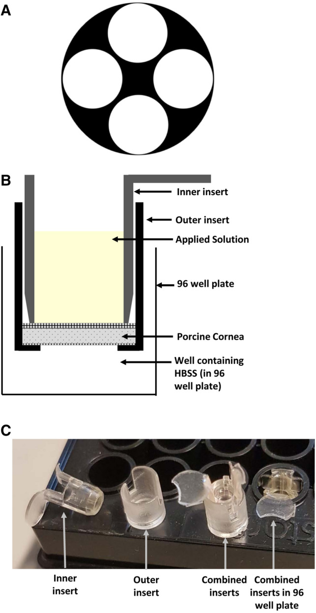
Figure 1.

Corneal model for measuring penetration. (A) Illustrative diagram of the location from which 5 mm discs were cut from the cornea. (B) Illustrative diagram showing a 5 mm porcine corneal disc fitted between plastic 96 well plate inserts. The combined insert is placed into a well containing Hanks’ Balanced Salt Solution (HBSS). Solution containing the compound of interest is then applied to the upper, epithelial, surface shown in yellow. (C) Photograph of inserts showing inner insert and outer inserts separately. The disc of corneal tissue is placed in the base of the outer insert and the inner insert placed on top to sandwich it against the base before placing the combined inserts into the 96 well plate.
