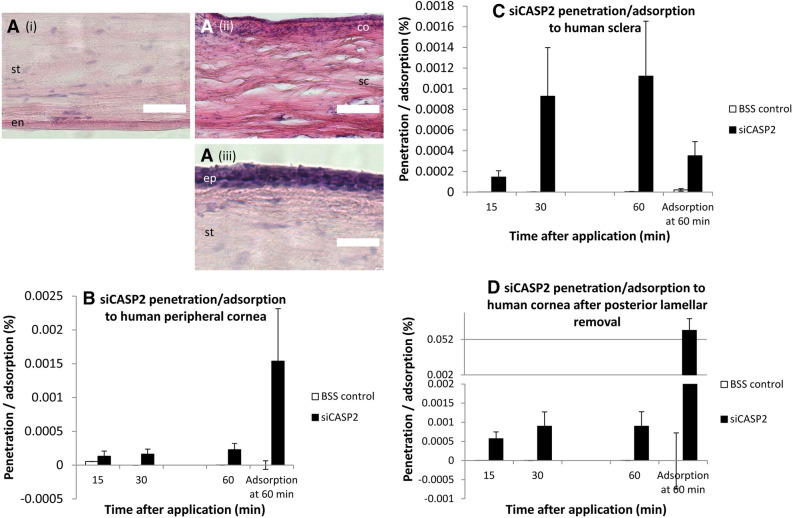
Figure 7.
(A) representative H&E-stained human corneal tissue from the assays in (B–D): (i), human peripheral cornea; (ii), human sclera; (iii), human central cornea after posterior lamellae removal. (B–D) Penetration of siCASP2 through and adsorption to human cornea and sclera over 60 min after siCASP2 was applied to the epithelial surface and washed off after 2 min. Results are displayed as bar charts with mean and standard error (error bars). (D) is displayed with a split y axis. “ep” corneal epithelium; “s” corneal stroma; “en” corneal endothelium; “sc” sclera; “co” conjunctiva; scale bar 100 µm. (B, C) represent data from n = 2 independent experiments; (D) represents data from n = 3 independent experiments.

