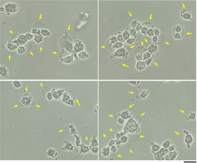
Figure 7.
The typical photos of the bubbles on the CAP-treated U87MG cells. Here, we showed four photos of the typical bubbling. A schematic illustration of the bubbling on the cell is shown in the middle. The photos were taken at 11 min post a 4 min of CAP treatment. The clear bubbles were marked by yellow arrows. 1.5 mL of U87MG cells (7.5 × 10⁴ cell/mL) were seeded in 35 mm dishes and cultured for 24 h in the incubator. The scale bar was 50 μm (black). In each row, the photos were taken in situ. All photos were taken by using a Nikon TS100 inverted phase-contrast microscope.

