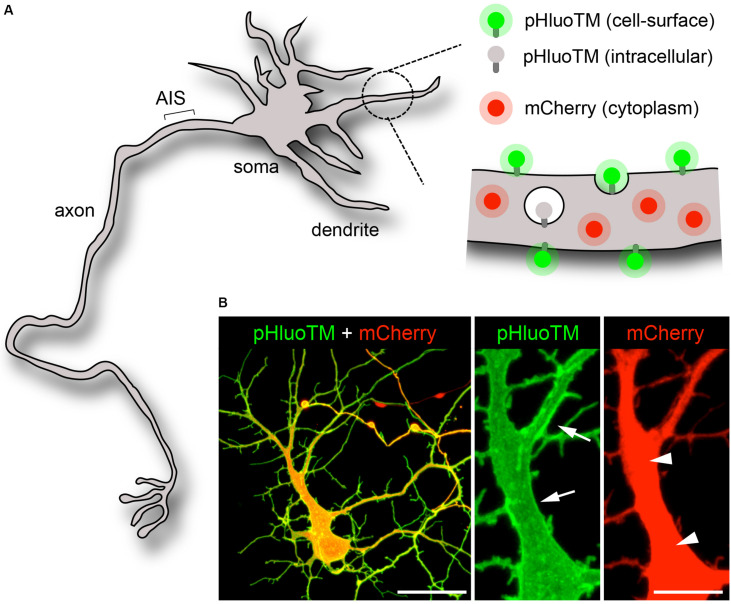
FIGURE 1.
Visualization of the neuronal cytoplasm and plasma membrane. (A,B) Scheme (A) and confocal pictures (B, maximal projections) of an immature neuron (DIV5) expressing pHluoTM (green) and mCherry (red). PHluoTM – a 39 KDa type I transmembrane protein derived from superecliptic (pH-sensitive) GFP – selectively fluoresces and marks the plasma membrane (arrows). mCherry, a 27KDa soluble protein, distributes throughout the cytoplasm (arrowheads). Scale bar 50 or 10 μm.

