FIGURE 6.
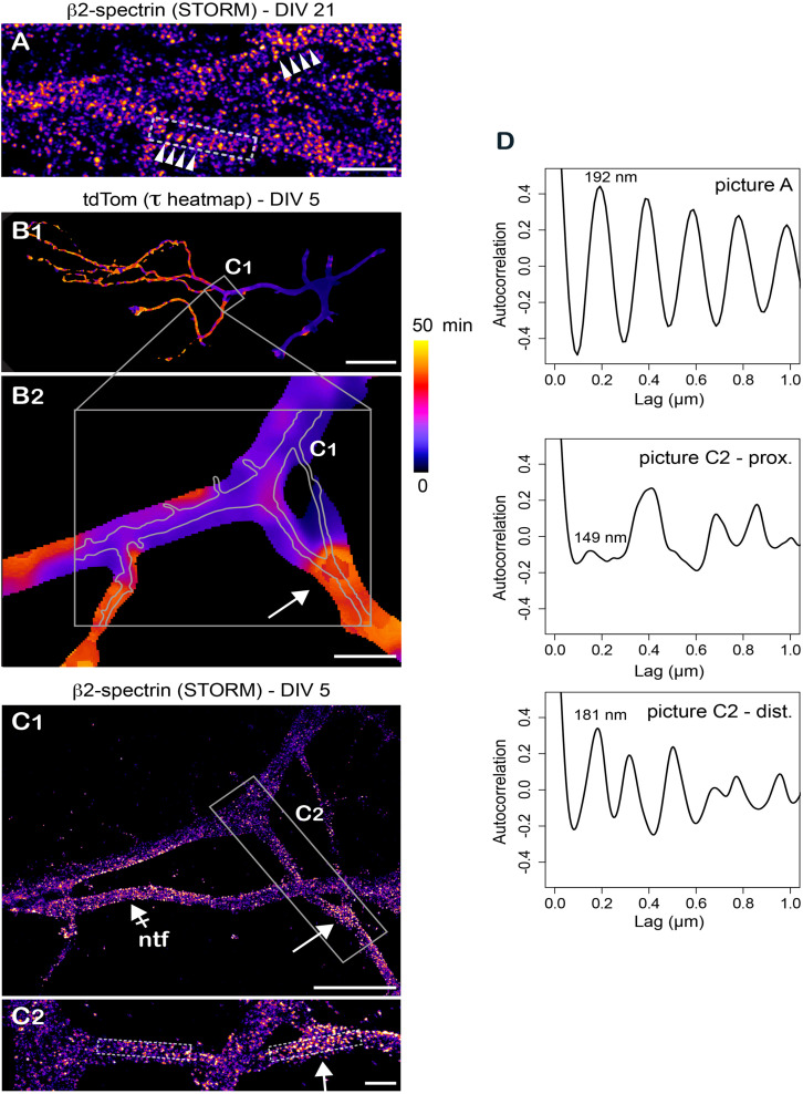
Retrospective stochastic optical reconstruction microscopy demonstrates the feasibility of correlative FLIP/super-resolution imaging. (A) Stochastic optical reconstruction microscopy (STORM) of βII-spectrin in axons of DIV 29 hipocampal neurons showing the repetitive organization of the subcortical cytoskeleton (spectin ring spacers, arrowheads). (B) Variation of tdTom diffusion throughout a DIV 5 neuron (tau heat maps) at low (B1) and higher (B2) magnification. Shown in gray in B2 are the position and contours of the axon imaged at higher resolution in (C). (C) Retrospective STORM imaging of βII-spectrin in the area highlighted in (B) at low (C1) and higher magnification (C2). Shown by a crossed-arrow in (C1) is a non-transfected neuron (ntf). In (B,C1,C2), note the presence of repetitive βII-spectrin structures in zones of transition from fast to slower diffusion of tdTom (arrows). (D) Auto-correlation signal along the longitudinal axes of areas highlighted in gray in (A,C2). For (C2), note the stronger periodicity of the signal in the distal dendritic segment and the strong resonance at 181nm in this segment. Scale bars 1, 50, 10, 5, and 1 μm in (A,B1,B2,C1,C2), respectively.

