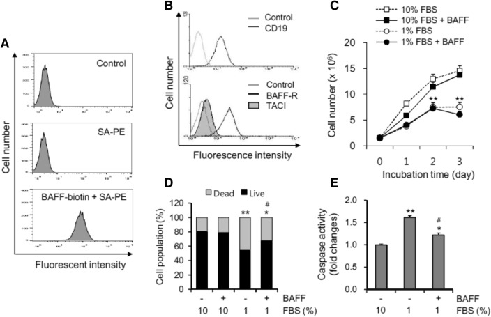
Figure 1.
Oxidative stress-induced cell death was inhibited by BAFF. (A) WiL2-NS cells were incubated with 10 μg/ml of a human BAFF-murine CD8 (BAFF-muCD8) biotinylated fusion protein on ice for 30 min. BAFF binding was visualized the incubation with phycoerythrin (PE)-conjugated streptavidin (SA) for 20 min and analyzed by flow cytometry. (B) WiL2-NS cells were incubated with biotinylated anti-human BAFF-R or TACI antibodies for 30 min followed by PE-conjugated SA for 20 min. At the same time, cells were incubated with FITC-labeled CD19. Expression of BAFF-R or TACI and CD19 on each cell was analyzed by flow cytometry as compared to control group incubated with isotype antibodies. (C) and (D) Cells were incubated in the RPMI 1640 medium with 10% or 1% FBS in the presence or absence of 20 ng/ml BAFF. Total number of cells (C) or dead cells (D) were counted with hemocytometer and estimated by trypan blue staining, respectively. Cell lysates were prepared and caspase 3 activity was measured by using Ac-DEVD-pNA, caspase 3 substrate. Caspase 3 activity was normalized with protein concentration (E). Data were the representative of four experiments. Data in the line (C) or bar (D–E) graph represent the means ± SD. *p < 0.05; **p < 0.01; significant difference as compared to BAFF-untreated control with 10% FBS (C, D and E). #p < 0.05; significant difference as compared to BAFF-untreated control with 1% FBS (D and E).

