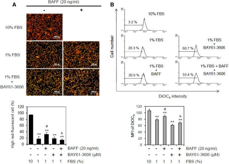
Figure 6.
Syk inhibitor enhanced MMP collapse by the incubation with 1% FBS. (A, B) WiL2-NS cells were incubated in the RPMI 1640 medium with 10% or 1% FBS in the presence or absence of BAY61-3606, Syk inhibitor and/or 20 ng/ml BAFF. Then, cells were stained with MitoProbe™ JC-1 reagent (A) or DiOC6 (B) for the detection of MMP. Cells were observed and photographed with 400 × magnification under fluorescence microscope (A, top). Number of high-red fluorescent cells was counted and represented as bar graph with mean ± SD (A, bottom). Percentage of cells with low fluorescence of DiOC6 was analyzed by flow cytometry (B, top). Mean fluorescence intensity (MFI) in each histogram was analyzed with software FlowJo V10. MFI was represented as bar graph with the means ± SD (B, bottom). All experiments were performed four times. **p < 0.01; significant difference as compared to BAFF-untreated and BAY61-3,606-untreated control with 10% FBS. #p < 0.05; significant difference as compared to BAFF-untreated and BAY61-3,606-untreated control with 1% FBS. &p < 0.05; significant difference as compared to BAFF-treated and BAY61-3,606-untreated control with 1% FBS.

