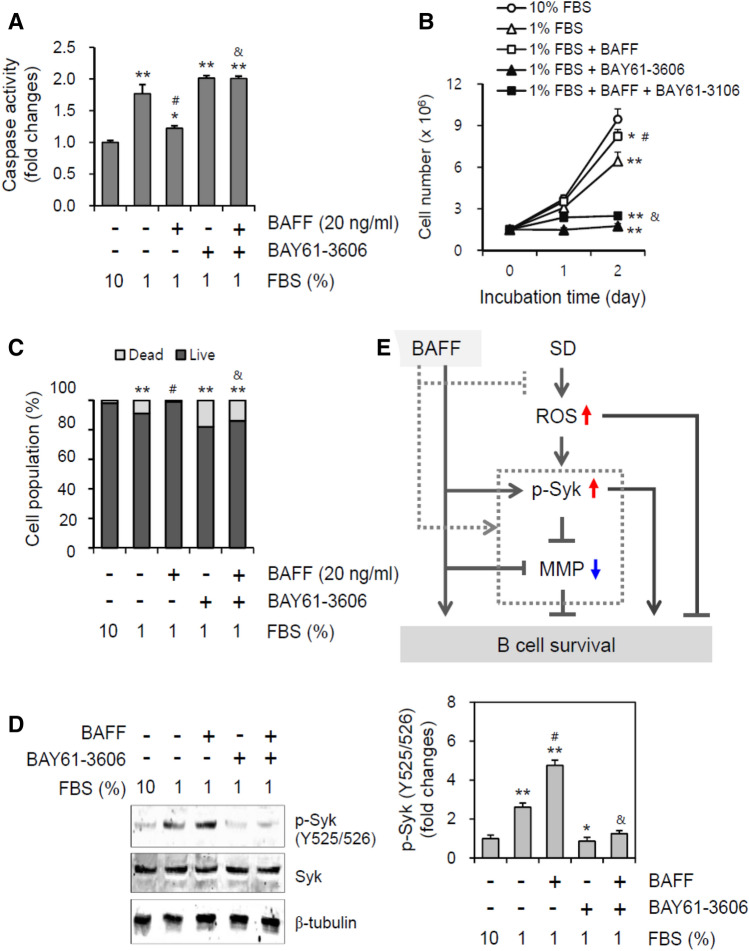
Figure 7.
Syk inhibitor increased B cell death by the incubation with 1% FBS. (A–D) WiL2-NS cells were incubated in the RPMI 1,640 medium with 10% or 1% FBS in the presence or absence of BAY61-3,606, Syk inhibitor and/or 20 ng/ml BAFF. Cell lysates were prepared and caspase 3 activity was measured by using Ac-DEVD-pNA, caspase 3 substrate. Caspase 3 activity was normalized with protein concentration (A). Total number of cells (B) or dead cells (C) were counted with hemocytometer and estimated by trypan blue staining, respectively. Cell lysates were prepared and western blotting was used to detect phosphorylated Syk at tyrosine (Y) 525/526. Processing (such as changing brightness and contrast) is applied equally to controls across the entire image (D, left). Each band for Syk phosphorylation at Y525/526 was quantified by using ImageJ 1.34 (D, right). All experiments were performed four times. Data in the line (B) or bar (A-D) graphs represent the means ± SD. *p < 0.05; **p < 0.01; significant difference as compared to BAFF-untreated control with 10% FBS. #p < 0.05; significant difference as compared to BAFF-untreated and BAY61-3,606-untreated control with 1% FBS. &p < 0.05; significant difference as compared to BAFF-treated and BAY61-3606-untreated control with 1% FBS (A, bottom). (E) This is the scheme for the regulation of serum deprivation (SD)-associated B cell survival via Syk-dependent mitochondria membrane potential (MMP). Our findings are indicated by gray-dotted lines. Black lines are from the results reported already in the literature.

