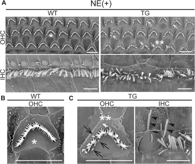
Fig. 3. Ultrastructural changes of stereocilia in DIA1-TG mice after noise exposure.
After noise exposure (NE) at the age of 4 weeks, the middle turns of cochleae in WT and DIA1TG/TG (TG) mice were fixed at the age of 8 weeks (at day 28 after NE) for scanning electron microscopy (SEM). Note that HC loss after NE was not significant in WT or TG mice. Scale bars: 5 μm. a Low-magnification views of OHCs (upper panels) and IHCs (lower panels) of the cochleae are shown. In the TG mice, stereocilia were damaged in some of the OHCs and IHCs. Abnormally short and sparse (arrows), and fused (arrowheads) stereocilia are indicated. b, c High magnification views of a surviving OHC in WT (b) and surviving OHC and IHC in TG mice (c). Asterisks and double asterisks indicate the same OHCs, while the IHC is from the adjacent portion of the same sample. The short (arrows) and elongated stereocilia (arrowheads) are indicated.

