. 2020 Jun;22(2):101–111. doi: 10.31887/DCNS.2020.22.2/mkorte

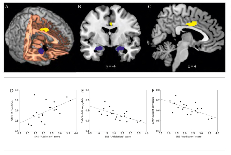
Figure 2. Magnetic resonance imaging of the human brain and analysis showing correlation between gray-matter 
volume (GMV) and social-networking site (SNS) addiction score. Depicted is the visualization of the voxel-wise–based 
morphometry (VBM) exemplified in three different views: (A) rendered brain; (B) coronal view; and (C) sagittal view. 
The SNS addiction score was negatively correlated with GMV in bilateral amygdala (shown as blue areas) and positively 
correlated with GMV in the anterior/mid cingulate cortex (ACC/MCC, shown as yellow area). Imaging is displayed in 
radiological view (right is on the viewer’s left). (D-F) Scatter plots show the pattern of correlation between GMV and SNS addiction score in (D) ACC/MCC, (E) left amygdala, and (F) right amygdala. Adapted from ref 57: He Q, Turel O, Bechara A. Brain anatomy alterations associated with Social Networking Site (SNS) addiction. Sci Rep. 2017;7:45064. doi:10.1038/srep45064. Copyright© 2017, The Authors.

Figure 2.