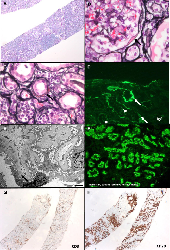
FIGURE 1.
Anti-LRP2 nephropathy with concurrent kidney infiltration by LPL (Case 1). (A) Kidney biopsy with diffuse lymphoplasmacytic inflammatory infiltrate (periodic acid–Schiff, ×50). (B) Immune deposit along Bowman’s capsule (Jones, ×200). (C) Partially circumferential TBM immune deposit; tubules have acute injurious changes with attenuation of their cytoplasm and surrounding lymphoplasmacytic inflammation (Jones, ×400). (D) Granular TBM (arrows) and focal tubular brush border (arrowheads) staining for IgG. (E) Segmentally distributed subepithelial immune deposits (transmission electron microscopy, direct magnification ×1900). (F) Indirect immunofluorescence of patient’s serum reacting with normal kidney tubular brush borders (image courtesy of Dr Chris Larsen, Arkana Labs). (G) Scattered CD3-positive T-cell infiltrate (×50). (H) CD20-positive B cells, compatible with involvement by LPL (50×).

