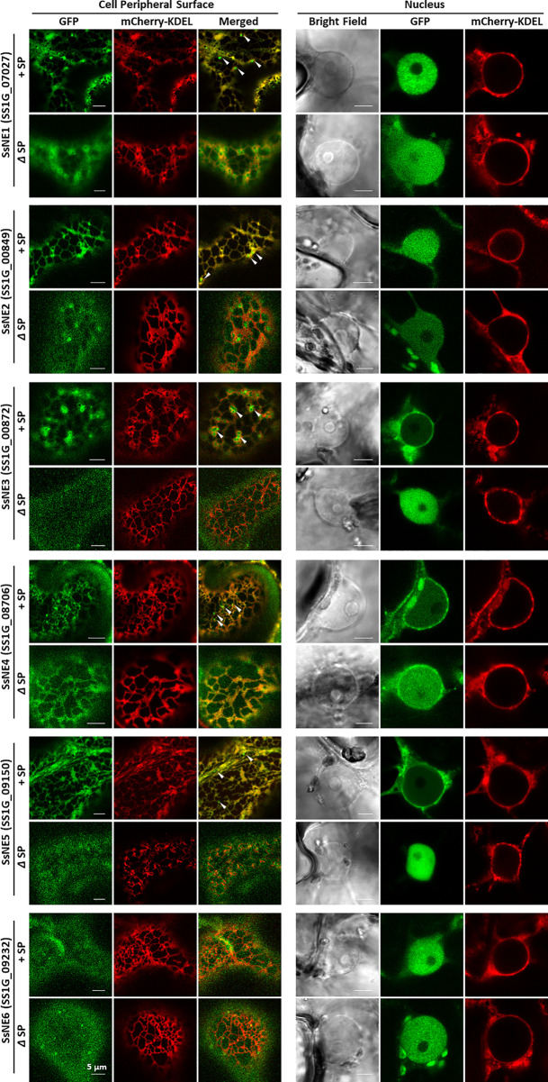
Figure 2.
Subcellular localization of Sclerotinia sclerotiorum necrosis-inducing effectors in Nicotiana benthamiana leaves. Each effector protein was fused to GFP at the C-terminus and tested with (+SP) and without its native signal peptide (ΔSP). All constructs expressing effector proteins were co-infiltrated into leaves with a construct expressing an mCherry protein tagged with an ER retention signal (mCherry::KDEL). Punctate signals adjacent to ER tubules that were not labeled by the mCherry-ER lumen marker are indicated with arrows.

