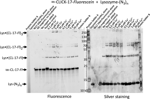
Figure 6.
SDS-PAGE gel showing Cu+- and Cu2+-dependence of in cis-catalyzed clicking of fluoresceinated ≡-CLICK-17 DNA to an azide-labeled protein (lysozyme-(N3)1–3). Both fluorescein fluorescence, indicative of DNA (left) and silver staining patterns for protein (right) are shown, both in grey scale. Lanes, from left to right, show Protein Ladder A; lysozyme-(N3)1–3 only; ≡-CLICK-17-Fluorescein only; lysozyme-(N3)1–3 mixed with ≡-CLICK-17-Fluorescein in the absence of either added copper or incubation; lysozyme-(N3)1–3 and ≡-CLICK-17-Fluorescein without added copper but incubated for 2.5 h; a positive control of the two reactants with 1 mM Cu+/5 mM THPTA for 2.5 h; the two reactants incubated for 2 h in the presence of 0.2 μM Cu+; 1 μM Cu+; 5 μM Cu+; 20 μM Cu+; 0.2 μM Cu2+; 1 μM Cu2+; 5 μM Cu2+; 20 μM Cu2+; and, Protein Ladder B.

