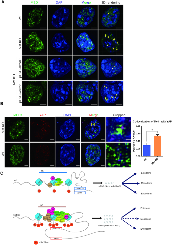
Figure 5.
YAP promotes Med1 condensates in Mst KO ESCs through phase separation. (A) Immunofluorescence pictures of MED1 puncta in WT and Mst KO ESCs as well as Mst KO ESCs with YAP and control knockdown taken by Nikon super-resolution microscope z-stack mode (3D-SIM). The nuclei were counterstained with DAPI. The rightmost 3D rendering pictures of Med1 phase separated condensates beyond unified criteria were rendered as yellow beads. The scale bars are 5 μm. (B) Immunofluorescence pictures of co-immunostaining of WT and Mst KO ESCs with MED1 and YAP antibodies. Pictures were taken by Nikon super-resolution microscope z-stack mode (3D-SIM). The nuclei were counterstained with DAPI. The rightmost cropped pictures show the area in yellow box with magnification. The scale bars are 5 μm and 1 μm in uncropped and cropped pictures respectively. Co-localization of channels for MED1 and YAP was quantified with Fiji Coloc 2 plugin. ROI (region of interest) is chosen in five different nuclei of WT or Mst KO ESCs. Average Pearson's R value is used to evaluate co-localization of two channels. (C) Model illustrating the mechanism of hyper-activated nuclear YAP in Mst KO ESCs induces preferential lineage differentiation through SEs.

