
Figure 3.
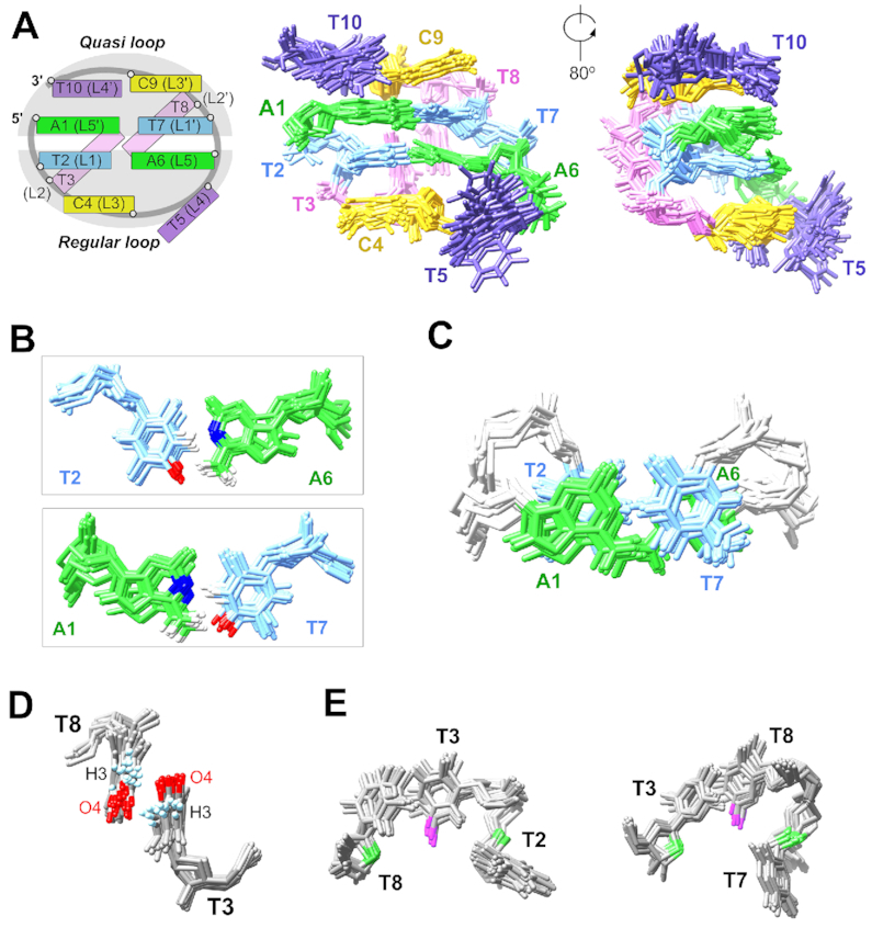
(A) Schematic of the ATTCT-Q/L5′ MDB and the superimposed 20 solution NMR structures (PDB ID: 6IY5). (B) T2-A6 and T7-A1 form Watson–Crick base pairs. (C) T2-A6 and T7-A1 base pairs show extensive base-base stackings. (D) T3 and T8 stack with each other, providing an orientation for favorable electrostatic interactions between T3 H3 and T8 O4, and between T3 O4 and T8 H3. (E) Dual hydrophobic cores formed by T3 methyl (magenta) and T2/T8 2′-methylene groups (green), and T8 methyl and T3/T7 2′-methylene groups.