Figure 7.

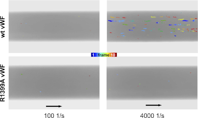
Binding of full-length vWF to ds DNA under shear flow conditions was captured by microfluidic experiments. Motion tracking of aggregates composed of ds DNA and wildtype vWF (upper row) and to vWF with the mutation R1399A in the A1 domain (lower row) is presented. Image compositions of 10 sequential frames each, taken at a frequency of 1.25 images per second, are shown highlighting the floating conglomerates in color. 60 s of high shear application of 4000 s−1 resulted in wt vWF-DNA conglomerates (upper right). In contrast, vWF–DNA interactions are not detectable neither for the vWF mutant R1399A nor for the case of the low shear stress of 100 s−1. Flow direction is indicated with black arrows corresponding to 100 μm.
