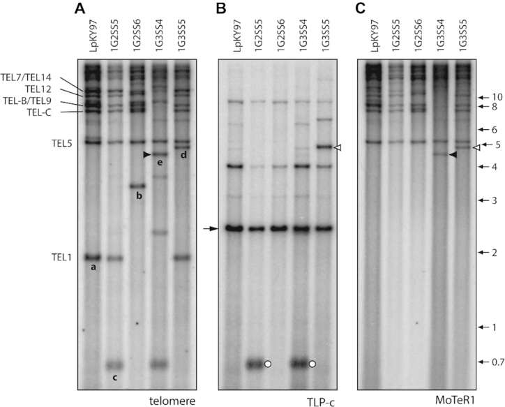
Figure 3.

Identification of putative MoTeR1 transposition events. The three panels show selected single spore DNA samples from the population shown in Figure 1. Panels A, B and C were hybridized with the telomere, TLP-c and MoTeR1 probes, respectively. (A) Novel TRFs that have been characterized are labeled ‘a’ through ‘e.’ (B) Re-probing of the blot with TLP-c produced a major signal in all lanes at a position corresponding to a molecular size of ∼2.5 kb. As expected, the probe hybridized to novel TRF-c, but also appeared to hybridize with novel TRF ‘d’ (open arrowheads). (C) The same blot was re-probed with the MoTeR1 probe. This also produced a hybridization signal at a position corresponding to TRF-d, implying that TRF-c had acquired a MoTeR1 insertion. Note that the MoTeR1 probe also hybridized to TRF-e (closed arrowheads)—signaling the occurrence of a second putative transposition event.
