
Figure 5.
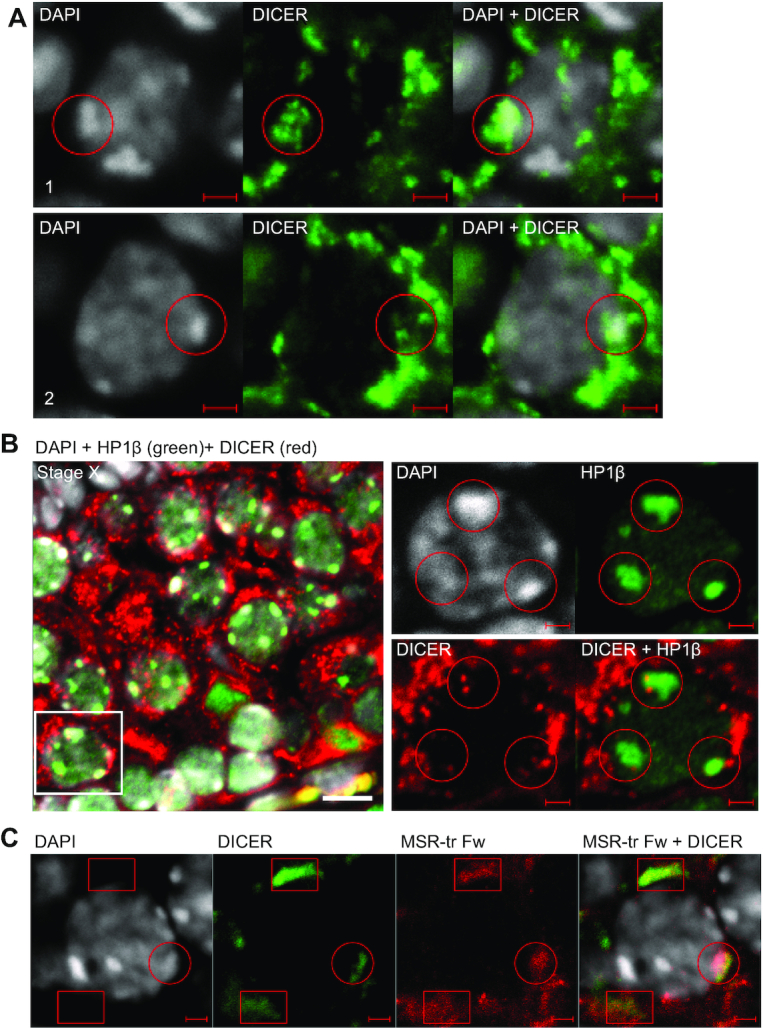
DICER localization in spermatocytes is associated with heterochromatin areas and MSR forward transcripts. (A) A panel shows a selected late pachytene spermatocyte from a PFA-fixed paraffin-embedded WT adult mouse testis section (stage IX) that was immunostained using an anti-DICER antibody (green). Nuclei were stained with DAPI (gray). Two different layers of the confocal Z-stack of the same cell are shown (1 and 2). DICER-positive foci are found in the nucleus, and the signal overlaps with DAPI-bright heterochromatin areas. Two different areas inside the same nucleus are indicated with red circles. Scale bars: 2 μm. (B) Nuclear DICER (red) is associated with heterochromatin as shown by co-immunostaining with anti-HP1β (green) on paraffin-embedded testis sections (stage X). Scale bar: 10 μm. Higher magnification images in the right panel show a single layer from a confocal stack to visualize an example spermatocyte indicated with a white box in the left panel. HP1β-positive heterochromatin areas are associated with DICER-positive foci (red circles). Scale bars in the high magnification images: 2 μm. (C) Immunofluorescence staining using an anti-DICER antibody (green) was combined with in situ hybridization using the MSR-tr Fw probe (red) on WT adult mouse testis cryosections. DICER partially co-localizes with MSR forward transcripts in the cytoplasm (red boxes) and in the nucleus (a red circle) of spermatocytes. Scale bars: 2 μm. All panels show a representative figure of an experiment that was independently repeated at least two times. See also Supplementary Figures S2–S4 and Supplementary Videos S1 and S2.