
Figure 3.
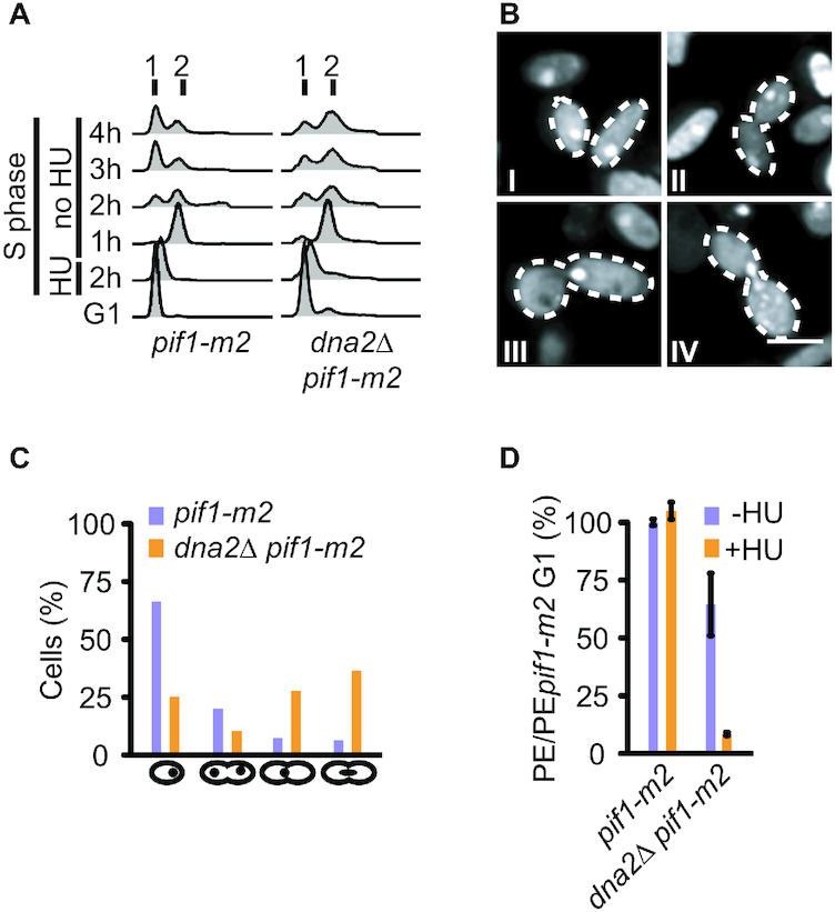
Transient RF arrest in the absence of Dna2 is cell-lethal. (A) Cell-cycle progression analysis by flow cytometry of the indicated strains synchronized in G1, treated with 200 mM HU for 2 h and released for 4 h into a drug-free medium. (B) Representative images of dna2Δ pif1-m2 cells treated as in panel A showing single-nucleated cells (I), a double-nucleated cell in G2 (II), a G2 cell with a nucleus at the bud neck (III), and an anaphase cell with an elongated nucleus spanning the bud neck (IV). Scale bar, 5 μm. (C) Quantification of cells (n ≥ 110 cells per strain) observed as in panel B. (D) Cell viability of the indicated strains, assessed by plating efficiency (PE) after synchronization in G1, removal of α-factor, and treatment or not with 200 mM HU for 2 h. Data expressed relative to pif1-m2 cells as mean values ± SD (n = 3 independent experiments).