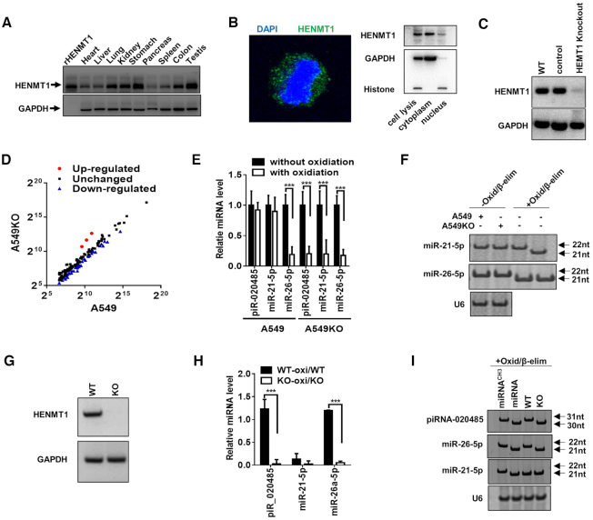
Figure 4.
HEMT1-knockout impairs 3′-terminal 2′Ome of miRNAs in A549 cells and mice. (A) HENMT1 expression in human tissues. (B) HENMT1 expression and location in A549 cell. Left: localization of HENMT1 detected by immunofluorescence analysis; right: localization of HENMT1 detected by Western blot. (C) HENMT1 expression detected by Western blot in WT A549 cells (WT), scramble gDNA (control cells) and HEMT1-knockout A549 cells (HEMT1 Knockout) via Crisper/cas9 technique. (D–F) Levels of miRNAs in A549 or HENMT1-knockout A549 (A549KO) cells detected by high-through sequencing after oxidation (D), qRT-PCR after oxidation (E) and Northern blot (F) after Oxid/β-elim procedure. (G) HENMT1 expression detected by western blot in lung derived from wild type mice (WT) and HEMT1-knockout mice (KO). (H) Ratio of piR_020485, miR-21-5p and miR-26a-5p levels with oxidation versus piR_020485, miR-21-5p and miR-26a-5p levels without oxidation in lung tissues from WT and HENMT1-KO (KO) mice by qRT-PCR. (I) Northern blot analysis of piR_020485 and miR-26a-5p expression in lung tissues from WT and KO mice after Oxid/β-elim procedure.

