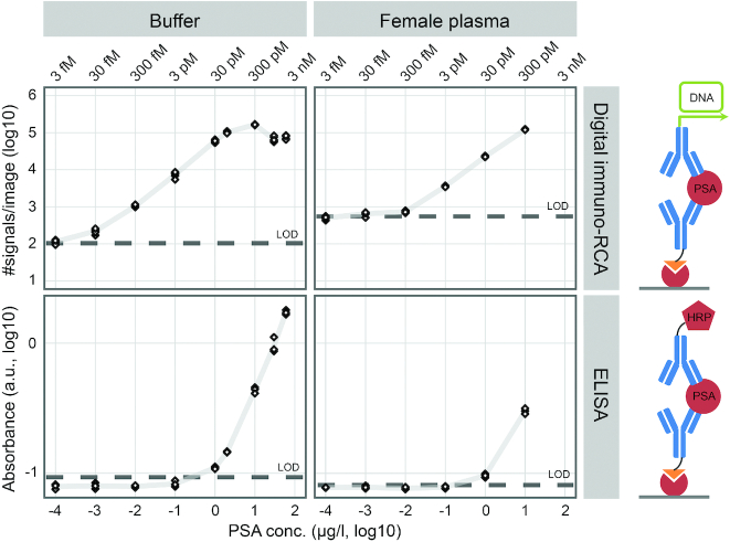
Figure 6.
Measurement of dilutions of PSA in buffer and female plasma with digital immuno-RCA and ELISA. The lowest PSA concentration prepared was 100 pg/l and the highest were 60 and 10 μg/l for buffer and female plasma, respectively. Negative controls were used to calculate LOD as three standard deviations above the mean. Six images from each sample were analyzed with digital immuno-RCA and triplicate samples were analyzed using ELISA. More than three orders of magnitude greater sensitivity was achieved when PSA was measured in buffer with digital immuno-RCA (∼100 pg/l) compared to ELISA (∼500 ng/l) (left panels). The difference in sensitivity was not as great for PSA spiked into female plasma, most likely due to low levels of endogenous PSA that could only be recorded using digital immuno-RCA. The same pair of monoclonal anti-PSA antibodies was used for both methods. The capture antibody was conjugated to biotin and the detection antibody was conjugated to DNA or HRP for digital immuno-RCA and ELISA, respectively. The percent values in the upper left panel represent detection efficiencies, i.e. the detected fraction of the added PSA molecules.

