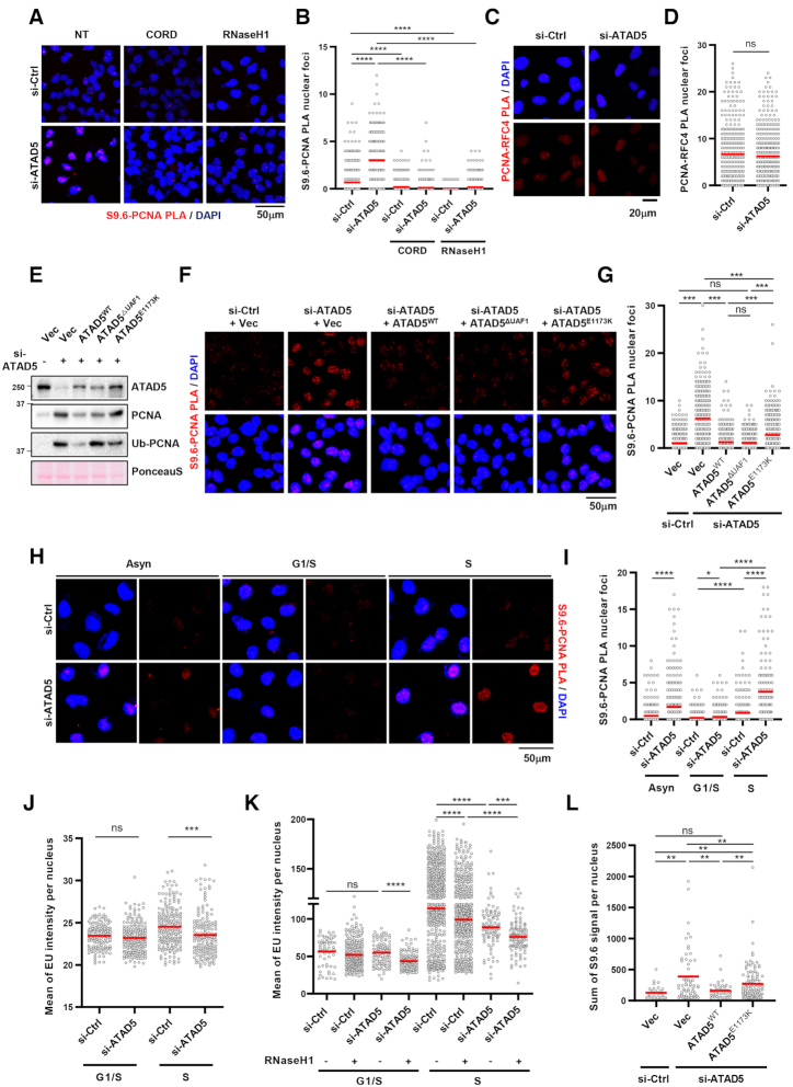
Figure 3.
PCNA remained on DNA by ATAD5 depletion generates R-loop signal. (A, B) After transfection and doxycycline treatment, HeLa-TetOn-RNaseH1-GFP cells were treated with 100 μM cordycepin for 2 h, fixed, and subjected to a PLA for S9.6 and PCNA. (A) Representative images. (B) Number of PLA nuclear foci was counted and plotted. (C, D) U2OS cells were transfected with ATAD5 siRNA for 48 h and fixed for a PLA between PCNA and RFC4. (C) Representative PLA images. (D) Number of PLA nuclear foci was counted and plotted. (E–G) U2OS-TetOn-ATAD5 cell lines were transfected with ATAD5 siRNAs and treated with doxycycline to express wild type ATAD5 (ATAD5WT), UAF1 interaction defective mutant ATAD5 (ATAD5ΔUAF1), or PCNA unloading defective mutant ATAD5 (ATAD5E1173K). After 48 h, chromatin-bound proteins were prepared for immunoblotting (E) or fixed for a PLA between S9.6 and PCNA (F, G). (F) Representative images of PLA. (G) Number of PLA nuclear foci was counted and plotted. (H–K) After siRNA transfection, cells were exposed to 2.5 mM thymidine for 18 h (G1/S boundary) and then released to fresh media for 5 h (S phase) before fixation. Asynchronous cells (Asyn) were also analyzed (H, I). (H–J) HeLa cells. (K) HeLa-TetOn-RNaseH1-GFP cells were transfected and treated with doxycycline for 48 h. Fixed cells were subjected to a S9.6-PCNA PLA (H, I) or EU-click analysis (J, K). (L) U2OS-TetOn-ATAD5 cell lines were transfected with ATAD5 siRNAs, treated with doxycycline to express ATAD5WT or ATAD5E1173K, and fixed for S9.6 immunostaining. (A–L) Three independent experiments were performed, and one representative result was displayed. Red bar indicates mean value. Statistical analysis: two-tailed Student's t-test; **** P < 0.001, *** P < 0.005, ** P<0.01, * P < 0.05 and n.s. not significant.

