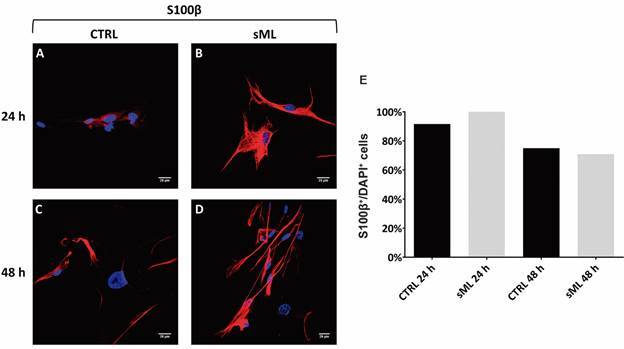
Fig. 1: immunophenotypic characterisation of human Schwann cells. Cells were obtained from healthy donors and cultivated for 24 and 48 h alone (CTRL) or in the presence of sonicated Mycobacterium leprae (sML). (A-D) Confocal microscopy showing immunodetection of S100β protein (red, AlexaFluor 594, Molecular Probes) and nuclear staining by DAPI (blue). Scale bar = 20 µm. (E) Graph shows the mean of the percentage of S100β+/DAPI+ cells.

