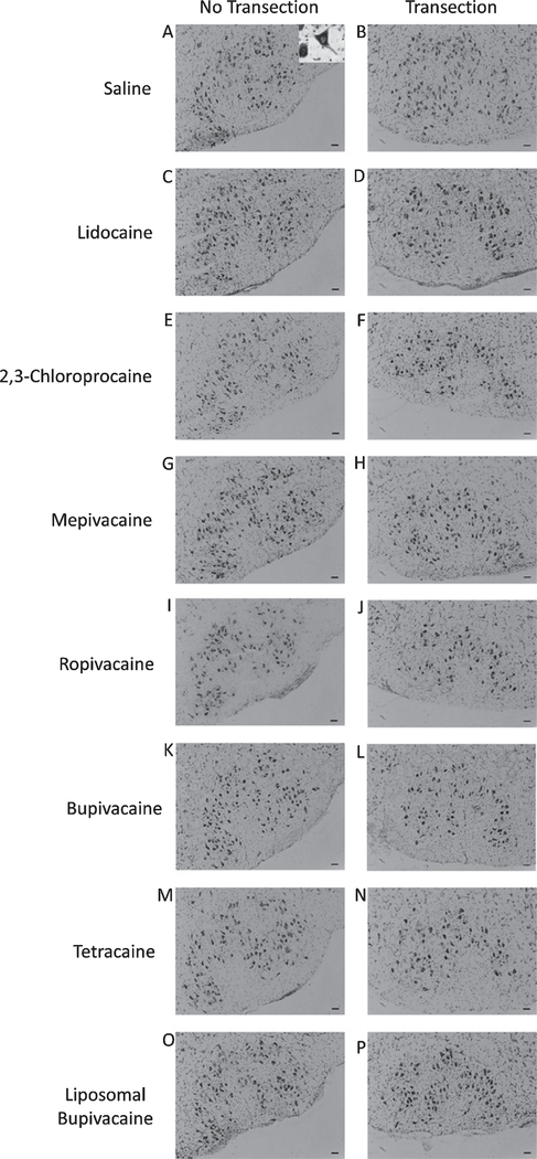
Fig. 1.
Thionin-stained facial motoneurons. Representative photomicrographs of thionin-stained facial motoneurons in the uninjured (no transection) and injured (transection) facial nuclei of mice treated with saline (A and B), lidocaine (C and D), 2,3chloroprocaine (E and F), mepivacaine (G and H), ropivacaine (I and J), bupivacaine (K and L), tetracaine (M and N), and liposomal bupivacaine (O and P) at the time of injury (immediate treatment). Scale bar - 50 μm. Inset of 1a is a magnified example motoneuron, demonstrating characteristic morphology and presence of distinct nucleus used for cell counting.

