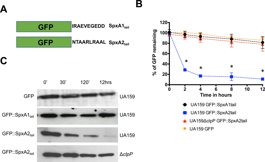
Figure 2.

Stability of GFP::SpxA1tail and GFP::SpxA2tail fusion proteins in S. mutans. (A) Graphical representation of GFP fused to the last 10 C-terminal amino acids of either SpxA1Smu or SpxA2Smu. (B) Fluorescence decay of GFP::SpxA1tail and GFP::SpxA2tail expressed in UA159 (parent) or ΔclpP strains after addition of chloramphenicol. Asterisks indicate time points showing statistically significant differences (p ≤ 0.01, one-way ANOVA) in decay of GFP expression in the SpxA2tail construct when hosted in UA159 compared to the ΔclpP strain. (C) Western blot analysis of UA159 or ΔclpP expressing GFP::SpxA1tail and GFP::SpxA2tail probed with anti-GFP polyclonal antibody. The images shown are representative of 3 or more independent experiments.
