
Figure 4.
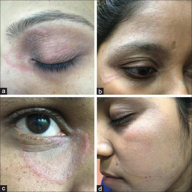
(a) Erythematous lesion of SD starting at the root of nose, involving both eyelids and ending at outer canthus. (b) “Double edged” lesion and powdery scales of tinea faciei visible on the right temple involving the entire upper eyelid and both canthi. (c) Erythematous scaly plaque with central clearing involving lower eyelid and inner canthus. (d) Fairly well defined powdery scaly lesion with mild erythema involving almost the entire left side of face including the eyelids