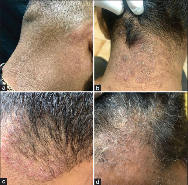
Figure 6.

(a) Well-defined lesion on the nape of the neck with fine scaling merging into the scalp. (b) Ill-defined erythematous lesion with scattered papules and scales merging into the scalp. (c) Well defined erythematous scaly papular eruption with a well-defined margin on the nape of necktraversing the scalp margin. (d) Tinea faciei on the forehead extending to the scalp
