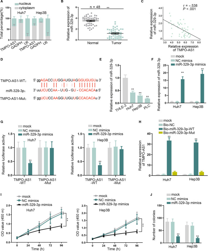
FIGURE 2.

MiR‐329‐3p acted as a target of TMPO‐AS1 and it negatively correlated with TMPO‐AS1 expression. A, The subcellular fraction assay was to test the RNA localization. B, MiR‐329‐3p expression was assessed in HCC tissues and normal liver tissues by qRT‐PCR. C, Pearson's correlation analysis was performed to analyze the correlation between miR‐329‐3p and TMPO‐AS1in HCC tissues. D, The binding site between miR‐329‐3p and TMPO‐AS1 was predicted by starBase. E, qRT‐PCR measured the expression of miR‐329‐3p in HCC cells and THLE‐3 cells. F, qRT‐PCR detected the transfection efficiency of miR‐329‐3p mimics. G, H, Luciferase reporter and RNA pull down assays confirmed the binding relation between miR‐329‐3p and TMPO‐AS1. I, J, CCK‐8 and colony formation assays tested cell proliferation under the effect of miR‐329‐3p mimics. ** P < .01
