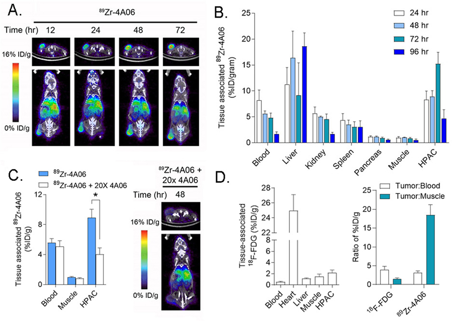
Figure 1. Detection of tumor autonomous expression of CDCP1 in a human pancreatic cancer model with 89Zr-4A06 PET/CT.
A. Representative transverse and coronal PET/CT images of a mouse bearing a subcutaneous HPAC xenograft at various time points after administration of 89Zr-4A06. The tumor is visible on the left hindlimb of the mouse. Some radiotracer accumulation is observed in normal abdominal tissues associated with immunoglobulin clearance, as expected. B. Ex vivo biodistribution data showing the accumulation of 89Zr-4A06 in mice bearing HPAC tumors over time. Tumor uptake of the radiotracer peaks at 72 hours post injection, while normal tissue accumulation is generally lower (n = 4/time point). C. At left are shown biodistribution data collected 48 hours post injection of 89Zr-4A06 or 89Zr-4A06 coadministered with 20x molar excess unlabeled 4A06 (n = 4/arm, *P<0.01). The excess antibody suppresses radiotracer binding in the tumor, as expected. At right are shown transverse and coronal PET/CT images of one mouse from the treatment arm that received 89Zr-4A06 with 20x unlabeled 4A06. D. At left are shown biodistribution data highlighting the uptake of 18F-fluorodeoxyglucose 60 minutes post injection in mice bearing subcutaneous HPAC tumors (n = 4). The level of uptake is lower than what was observed for 89Zr-4A06. Selected normal tissues are shown for comparison. At right are shown a comparison of the tumor to blood and tumor to muscle ratios for 18F-FDG (60 min post injection) and 89Zr-4A06 (72 hours post injection).

