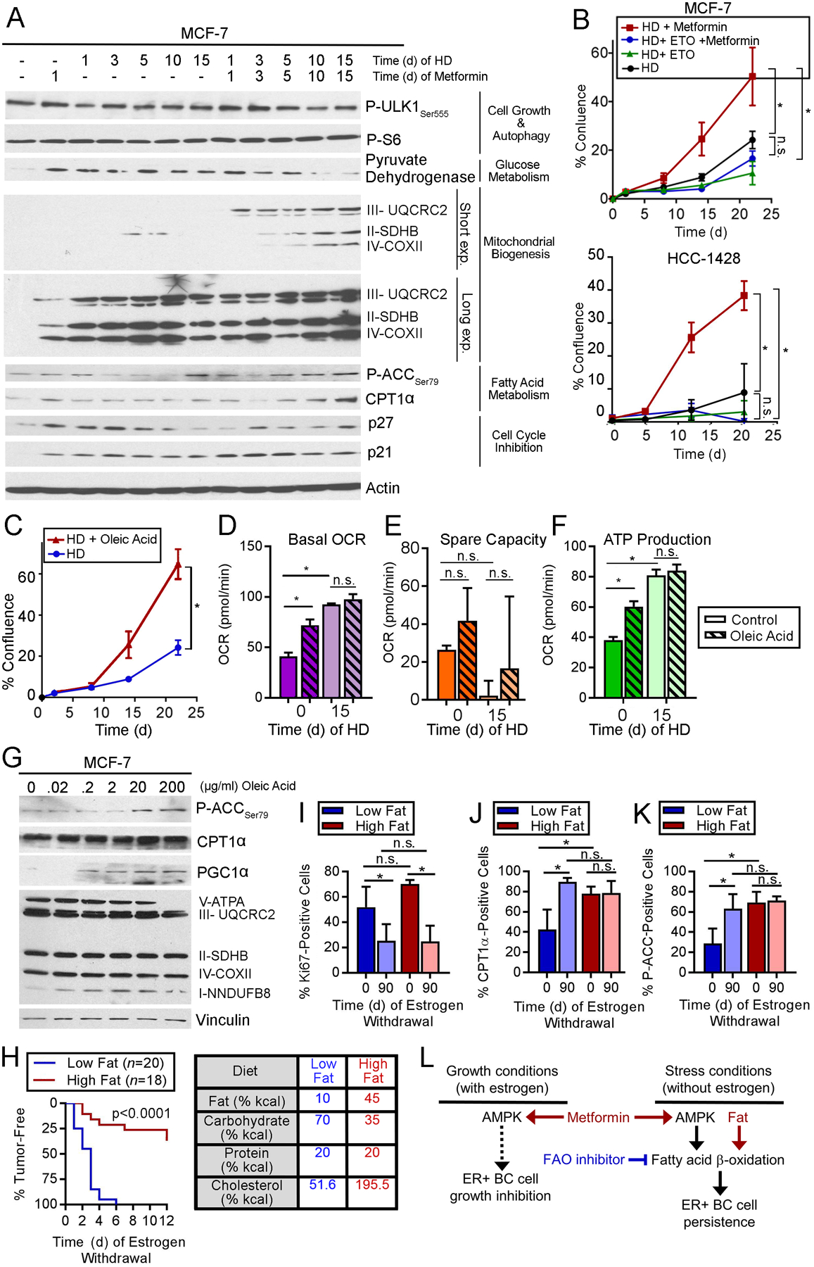
Fig. 6. Dietary fat promotes survival of ER+ BC cells following estrogen withdrawal.

(A) Immunoblot analysis of lysates from cells treated with HD ±1 nM E2 and metformin. (B/C) Cells were treated with HD ±200 μM etomoxir or 1 mM metformin (B), or ±300 μg/mL oleic acid (C). Relative viable cells were serially measured. (D-F) OCR was measured from cells treated with HD ±1 nM E2 for 15 d, then treated ±200 μg/mL oleic acid for 5 h. Basal OCR (D), spare capacity (E), and ATP production (F) were measured. (G) Lysates of cells treated ± oleic acid for 24 h were analyzed by immunoblot. (H) Ovx mice pre-conditioned to either high-fat or low-fat diet for 2 wk were injected orthotopically with MCF-7 cells and supplemented with E2 (with diet continuation). When tumors reached ~400 mm3, E2 was withdrawn. Tumor volumes were serially measured. Data are presented as proportions of mice reaching complete tumor regression over time. Groups were compared by log-rank test. Caloric sources in diets are noted at right. (I-K) IHC for Ki67, CPT1α, or P-ACCSer79 using tumors harvested after 0 or 90 d of EW. (L) Model summarizing observations of effects of AMPK and FAO on ER+ BC cells and tumors. In (B/C), *p≤0.05 by repeated measures ANOVA. In (D-F) and (I-J), *p≤0.05 by Tukey’s multiple comparison-adjusted posthoc test vs. baseline unless otherwise indicated. n.s.-not significant. In (B-F) and (I-J), data are shown as mean of triplicates ± SD.
