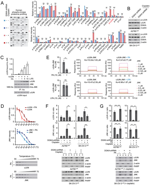
Figure 3. DGKA and PA activate c-JUN through c-JUN-JNK complex formation and nuclear localization upon cisplatin treatment.
A, Phospho-kinase proteome profiling of A2780cisR cells with DGKA knockdown and cisplatin treatment. Cells were treated with a sublethal dose of cisplatin for 48 hr. Density analysis was performed using ImageJ software. B, Phospho-cJUN Ser63 levels in A2780cisR and SK-OV-3cisR cells treated with DGKA shRNA and cisplatin. C, Effect of PA on c-JUN and JNK interaction. Recombinant JNK and c-JUN were incubated with increasing concentrations of PA. D, Cellular thermal shift assay (CETSA) curves for c-JUN or JNK measured in cell lysates harboring myc or flag tagged c-JUN or JNK treated with or without 10 μM PA. E, SPR analysis of interaction between c-JUN and JNK in the presence of PA or LPA. F and G, Localization of c-JUN and JNK in cells with DGKA knockdown and cisplatin treatment in (F) and PA rescue in (G). β-actin and PARP were used as cytoplasmic and nuclear markers, respectively. c: cytosol, n: nucleus. (D) n=3 and (A) n=2 technical replicates. Results of one representative experiment from four (C and D), three (B, F and G), two (E) and one (A) independent experiments are shown. Error bars represent SD. P values were determined by one-way ANOVA (ns: not significant; *P < 0.05; **P < 0.01; ***P < 0.001; ****P < 0.0001).

