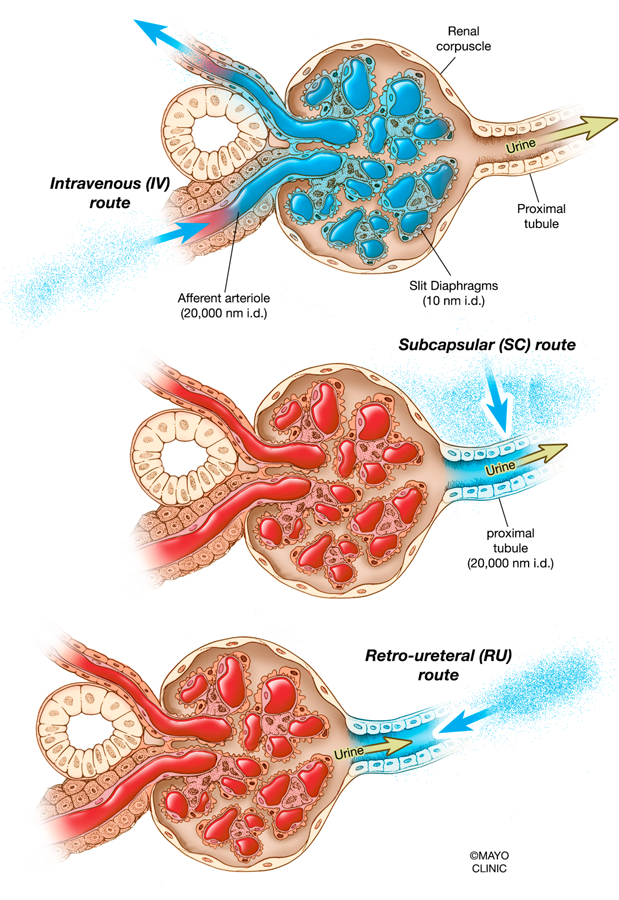
Figure 2. Intravenous and direct kidney vector administration in the context of the renal corpuscle.

Injecting vectors by the intravenous route would result in most vectors being stuck in the glomeruli of the kidneys or recirculated into the bloodstream, since slit diaphragms are approximately 10 nm in diameter (upper). Injecting vectors by the subcapsular route would bypass the glomeruli and potentially give vectors access to the apical and basolateral sides of some tubules (middle). Injecting vectors by the retro-ureteral route would theoretically grant vectors access to the apical side of tubules unless they escape from within the tubule epithelium, since proximal tubules are up to 20,000 nm in diameter (lower). nm, nanometer; i.d., in diameter.
