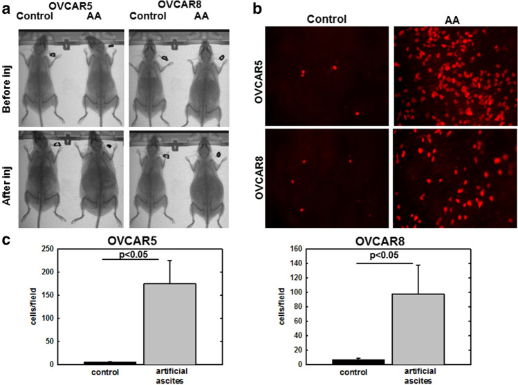
Figure 1.
Artificial ascites model of compression enhances OvCa cell adhesion to peritoneum in vivo. (A) MicroCT Scans showing C57Bl/6 female mice injected i.p. with 1 mL (control) or 5 mL (artificial ascites, or AA) PBS containing 106 RFP-tagged OVCAR5 or OVCAR8 cells, as indicated, for 5 or 8 h, respectively. (B) Mice were sacrificed, peritoneal tissue was collected and images of adherent cells to peritoneum were obtained using Echo Revolve fluorescent microscope at × 20 magnification. (C) Adherent cells were quantified using ImageJ. All experiments in were performed as triplicates with three independent biological replicates per cell line. All results are presented as mean ± s.e.m. and P-values were calculated using a Student’s two-tailed t-test. P < 0.05 is statistically significant.

