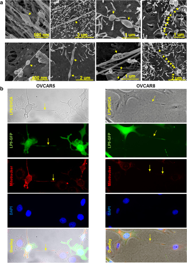
Figure 6.
Compression-induced nanotubes adopt distinct morphologies and participate in mitochondria transport between LP9 mesothelial cells and OvCa cells. (A) High magnification scanning electron micrographs of TNT formed under compression (~ 3 kPa; ~ 22 mmHg) in murine peritoneal explants. The peritoneal explants were compressed ex vivo for 1 h, fixed and processed for imaging using a FEI-Magellan 400 Field Emission Scanning Electron Microscope. Yellow arrows refer to distensions in the nanotubes. (B) GFP-tagged LP9 human peritoneal mesothelial cells were incubated with MitoTracker red to label mitochondria, then co-cultured with OVCAR5 or OVCAR8 cells under compression (~ 3 kPa; ~ 22 mmHg) for 24 h and stained with DAPI. Cells were imaged with Leica DM5500 fluorescence microscope at × 20 magnification. Top panel: phase image showing LP9 and OVCAR cells; second panel: GFP-tagged LP9 peritoneal MC; third panel: MitoTracker red showing labeled mitochondria in LP9 cells and extracellular projections; fourth panel: DAPI-stained nuclei of both LP9 and OVCAR cells; fifth panel: overlay. Yellow arrows denote nanotubes or labeled mitochondria transferred in nanotubes.

