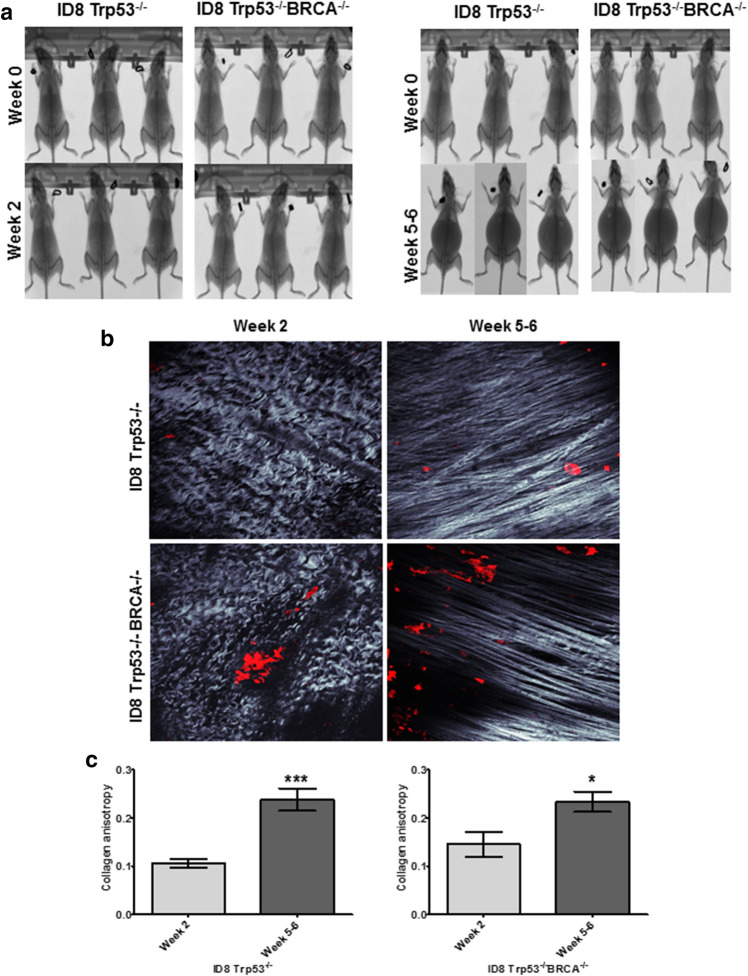
Figure 7.
Ascites accumulation in vivo correlates with enhanced collagen anisotropy. (A) C57Bl/6 female mice were injected i.p. with 5 × 10⁶ RFP-tagged ID8-Trp53−/− or ID8-Trp53−/− BRCA−/− syngeneic murine OvCa cells as indicated. MicroCT images were procured at time of injection (week 0) and time of sacrifice (week 2 or week 5–6 post-injection, as indicated). (B) The parietal peritoneum was dissected and prepared for combined fluorescence/SHG microscopy as described in Methods. Shown are representative examples from each cohort to show collagen fiber alignment (grey) and metastatic lesions (RFP-tagged cancer cells, red). (C) Collagen anisotropy was quantified using ImageJ. All results are presented as mean ± s.e.m. and P-values were calculated using a Student’s two-tailed t-test.

