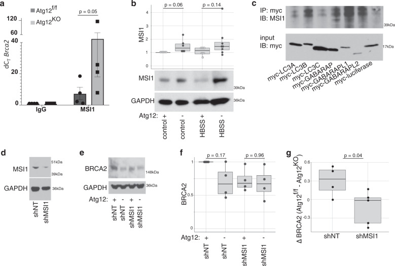
Fig. 7. Increased MSI1 in autophagy-deficient cells impairs Brca2 translation efficiency.
a Quantification (mean ± SD, n = 4 biologically independent replicates) of the fold enrichment of Brca2 interaction with MSI1 over IgG control in Atg12f/f or Atg12KO MEFs by RNA immunoprecipitation. An outlier (Dixon test p = 0.007) was excluded from statistical analysis, and the p value by t test is shown. b Boxplot, with dotplot overlay for each biological replicate, of relative MSI1 protein levels normalized to loading control and representative immunoblots from autophagy-inhibited MEFs, assayed by immunoblotting. Statistical analysis was performed by t test. c Representative immunoblot of immunoprecipitation of myc-tagged overexpressed LC3 family members interaction with MSI1 in HEK293T cells. Immunoprecipitation was performed with three biologically independent replicates. d Protein lysate was collected from Atg12f/f MEFs treated with shRNA to Msi1, and immunoblotted as indicated. Knockdown of ~50% was consistent among three independent biological replicates. e–g Protein lysate was collected from Atg12f/f MEFs that were stably knocked down for MSI1 and subsequently treated with 4OHT or control, and assayed by immunoblotting. e Representative immunoblot is shown. f Quantification of relative BRCA2 protein levels normalized to loading control, are plotted as a boxplot with dotplot overlay for each independent biological replicate, p values by ANOVA with Tukey’s post hoc test. g Difference in BRCA2 steady state protein levels (ΔBRCA) between Atg12f/f and Atg12KO MEFs following MS1 depletion, p value by t test.

