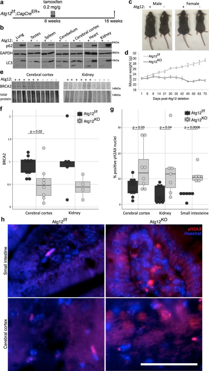
Fig. 9. Atg12 deletion in vivo leads to reduced BRCA2 and increased DNA damage.
a Diagram of Atg12f/f;Cag-CreER+ mouse treatment and tissue collection. b Protein lysate was collected from tissues two weeks following vehicle or 0.2 mg/g tamoxifen treatment and immunoblotted for markers of autophagic flux (p62/SQSTM1, LC3). c Representative images of male and female Atg12f/f and Atg12KO littermates. d Body weights of male mice following Atg12 deletion (Atg12f/fn = 17, Atg12KOn = 14). e Protein lysate was collected from mouse tissues 10 weeks following vehicle or tamoxifen treatment, and immunoblotted for BRCA2. f Boxplot with dotplot overlay for biological replicates of BRCA2 protein levels, normalized to total protein levels, assayed by immunoblotting. p = 0.02 by t test. g Boxplot with dotplot overlay for biological replicates of percent of γH2AX-positive nuclei by immunofluorescence, counted over four randomly selected fields of stained tissue per a minimum of four mice. p value calculated by t test. h Representative immunofluorescence for γH2AX (red) in mouse tissues from the cerebral cortex and small intestine, with nuclei counterstained by Hoechst (blue). Bar = 50 μm.

