
Figure 4.
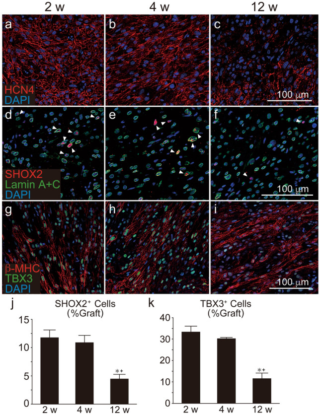
Chronological expression of nodal markers in grafted human ES cell-derived cardiomyocytes (hESC-CMs) in injured hearts. hESC-CMs were transplanted on week after induction of myocardial infarction. Histological analysis was performed at the same time-points. (a–c) HCN4 (red) staining in the graft area. (d–f) SHOX2 (red) and human specific Lamin A+C (green) staining. SHOX2+/Lamin A+C+ cells (arrow heads) were designated as graft nodal cells. (g–i) Staining against β-MHC (red) and TBX3 (green). (j) Quantitative analysis of the number of SHOX2+/Lamin A+C+ cells divided by the number of total Lamin A+C+ graft cells (n = 5 per group). Data represent mean ± SEM. *P = 0.0036 vs. 2 weeks, + P = 0.0116 vs. 4 weeks by ANOVA with Tukey’s post hoc test. (k) Quantitative analysis of the number of TBX3 + cells divided by that of β-MHC+ graft cardiomyocytes (n = 5 per group). Data represent mean ± SEM. *P = 0.0003 vs. 2 weeks, + P = 0.0015 vs. 4 weeks by ANOVA with Tukey’s post hoc test.