
Figure 2.
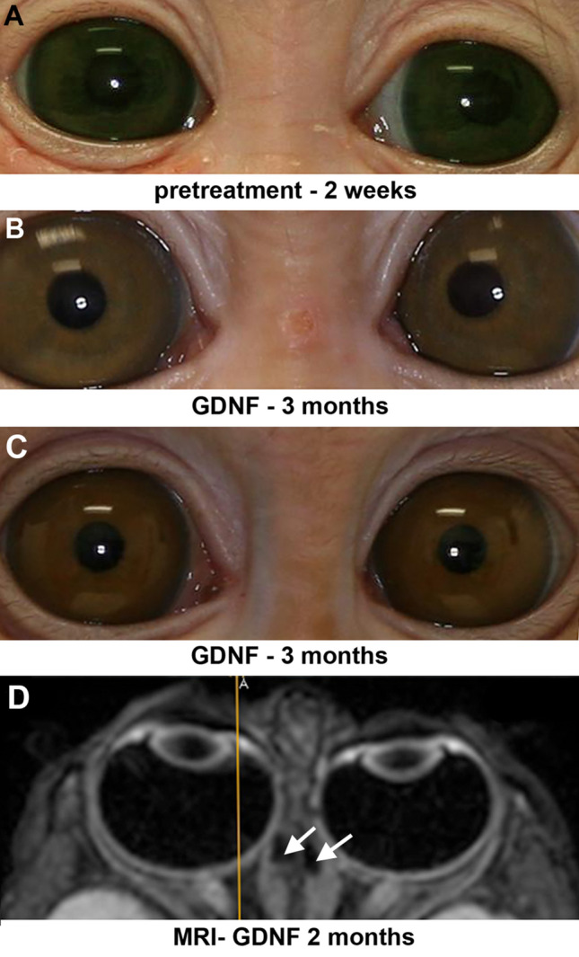
Photomicrographs of monkey 1 prior to treatment at 2 weeks of age (A) and at the end of 3 months of GDNF treatment for the one monkey that developed esotropia (B), and for one example of one of the monkeys that developed exotropia (M2) (C). (D) Magnetic resonance imaging (MRI) image of monkey 3 two months after implantation of a GDNF-releasing pellet on the right medial rectus muscle and a placebo control pellet on the left medial rectus muscle. The yellow line was generated by the MRI image acquisition system to indicate orientation. A indicates anterior. All monkeys in this group were male. Arrows indicate pellets on the medial rectus muscles.